If you have well-controlled diabetes, all-on-x dental implants can be a safe and effective way to replace missing teeth and preserve jawbone — with good glycemic control and committed aftercare, your risk of complications is similar to that of non-diabetic patients. This article shows how diabetes affects oral and bone health, how clinicians assess eligibility, and what to expect during surgery and recovery.
You'll learn practical steps to prepare for implants, the tests and planning clinicians use to lower risk, and the long-term factors that influence success so you can make an informed decision about your treatment.
Table of Contents
ToggleImpact of Diabetes on Oral and Bone Health
Diabetes affects healing, bone metabolism, and infection risk in measurable ways that influence implant planning and maintenance. Pay attention to blood glucose levels, bone quality at the implant site, and periodontal status to reduce complication risk.
Glycemic Control and Oral Healing
Poor glycemic control slows soft-tissue healing and increases infection risk after surgery. Hyperglycemia impairs neutrophil function and collagen synthesis, so elevated HbA1c (commonly >7% or individualized by your provider) correlates with higher early implant complications.
If your diabetes is well controlled, you generally experience healing rates closer to non-diabetic patients. You should still plan for longer soft-tissue maturation and schedule follow-ups at shorter intervals in the early postoperative period.
Preoperative optimization—short-term glycemic stabilization, coordinating with your physician, and perioperative antibiotic strategies when indicated—reduces risk. Monitor wound sites for delayed closure, excessive inflammation, or pus, and act promptly if these signs appear.
Bone Density Considerations
Diabetes alters bone remodeling through changes in osteoblast and osteoclast activity and advanced glycation end-products that stiffen collagen. These changes can reduce local bone turnover and may lower primary stability in some cases, especially in long-standing or poorly controlled diabetes.
You should obtain high-quality radiographic assessment (CBCT when indicated) to evaluate cortical thickness and trabecular density at the planned implant site. Consider surgical modifications—undersized osteotomy, staged approach, or use of wider/longer implants—and adjuncts like bone grafting or growth factors when native bone appears compromised.
Assess systemic factors that affect bone (vitamin D status, smoking, medications like bisphosphonates) because they interact with diabetic bone changes and influence implant osseointegration and long-term support.
Periodontal Health in Diabetic Patients
Diabetes increases the prevalence and severity of periodontitis through amplified inflammatory responses and impaired host defense. You are at higher risk for deeper pockets, greater attachment loss, and faster progression if glycemic control is suboptimal.
Control of periodontal disease before implant placement is essential. Perform thorough debridement, treat active infection, and confirm stable periodontal status with probing and radiographs.
After implant placement, maintain a strict hygiene regimen and schedule more frequent maintenance visits (often every 3–4 months). Early detection and management of peri-implant mucositis prevents progression to peri-implantitis, which has higher incidence and worse outcomes in patients with poor glycemic control.
Eligibility and Risk Assessment
You need clear criteria for implant candidacy and a focused review of health factors that affect healing and infection risk. The following subsections cover blood sugar control, relevant medical history, and specific contraindications or precautions to consider.
Assessing Glycemic Stability
Measure your HbA1c within three months before implant surgery; many clinicians use a target of <7.0% for routine implant placement. If your HbA1c falls between 7.0% and 8.5%, discuss risks with your dentist and physician; some teams will proceed with enhanced perioperative controls, prophylactic antibiotics, and more frequent follow-up.
Also evaluate fasting glucose and daily glucose logs if you use insulin or have large glycemic variability. Document recent hypoglycemia or hyperglycemia episodes because wide swings increase infection and healing complications. Plan to optimize control for at least 6–8 weeks before surgery when possible, and arrange closer postoperative glycemic monitoring during the first 2–4 weeks of healing.
Medical History Evaluation
Provide a current list of medications, including insulin, oral hypoglycemics, antiplatelets, and immunosuppressants. Steroids, biologic agents, and poorly controlled cardiovascular disease alter risk and may require consultation with your physician.
Assess renal function, hepatic status, and any history of radiation or chemotherapy to the head and neck. Review smoking status and periodontal disease history; both substantially raise peri-implantitis and failure risk. Document wound-healing disorders, neuropathy, and vascular disease, since peripheral vascular disease correlates with slower osseointegration.
Contraindications and Precautions
Absolute contraindications include active, uncontrolled systemic infection and untreated oral infections such as severe periodontitis. Major uncontrolled comorbidities—unstable cardiac disease, recent myocardial infarction, or uncontrolled hypertension—should delay elective implant surgery.
Exercise caution with poor glycemic control (HbA1c consistently >8.5%), heavy smoking (>10 cigarettes/day), or active bisphosphonate/antiresorptive therapy without specialist clearance. Implement precautions: preoperative antibiotic prophylaxis when indicated, staged implant placement for high-risk sites, and an individualized maintenance schedule with professional cleanings every 3–4 months for the first year.
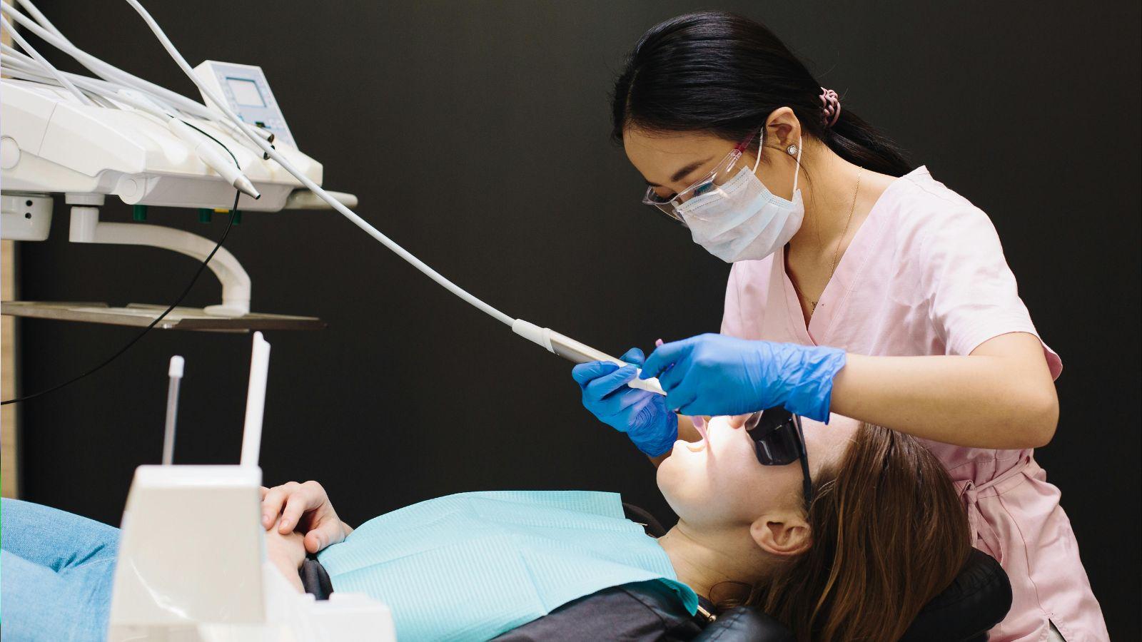
Surgical Planning and Post-Operative Care
You will need precise pre-surgical checks, careful selection of prosthetic materials, targeted infection-prevention measures, and a structured maintenance plan to optimize implant outcomes when your diabetes is controlled. Each step reduces risk and supports long-term bone and soft-tissue health.
Pre-Surgical Preparation
Confirm HbA1c within the target range your clinician set—typically ≤7% for most patients—within 3 months before surgery. Review medications that affect healing (antiplatelets, steroids) and coordinate adjustments with your physician.
Optimize oral health: eliminate active periodontal disease and treat caries before implant placement. Schedule scaling and root planing or extractions at least 4–6 weeks prior to allow soft-tissue healing and reduce bacterial load.
Plan implant positioning with CBCT and digital guides to avoid thin cortical plates and to maximize primary stability. Aim for insertion torque and implant length that achieve primary stability without overcompression of bone.
Arrange perioperative glycemic control: schedule morning procedures after light meals and insulin adjustments as advised. Have glucose monitoring available on the day of surgery and a plan for hypoglycemia or hyperglycemia management.
Prosthetic Materials Selection
Choose implant-abutment connections and materials that minimize microgap and bacterial leakage; internal conical connections reduce micromovement risk. Use titanium or titanium alloys for fixtures; they balance osseointegration and mechanical strength.
Select abutment and crown materials based on load, esthetics, and plaque resistance. Zirconia crowns offer low plaque affinity and good esthetics in the anterior; high-strength ceramic or metal-ceramic options suit posterior load-bearing restorations.
Consider overdentures on two implants with resilient attachments when bone quantity is limited; they simplify hygiene for patients with dexterity or glycemic-related neuropathy. Use screw-retained prostheses where possible to facilitate removal for hygiene or peri-implantitis management.
Verify occlusion and reduce lateral forces with splinting or adjusted contours. Provide clear written instructions on cleaning around specific materials and components to prevent plaque accumulation.
Infection Prevention Strategies
Administer prophylactic antibiotics based on your clinician’s protocol and medical history; many teams use a single preoperative dose for patients with well-controlled diabetes. Discuss allergy and prior antibiotic exposure with your provider.
Adopt strict aseptic technique in the operatory: sterile fields, minimal flap elevation when appropriate, and atraumatic handling of soft tissue. Limit surgery time to reduce exposure and tissue desiccation.
Use chlorhexidine 0.12–0.2% mouthrinse starting the evening before and for 1–2 weeks post-op as directed. Provide topical antiseptic instructions and reinforce smoking cessation to lower infection risk.
Plan early postoperative reviews at 1 week and again at 2–4 weeks to detect infection signs (swelling, purulence, persistent pain). Have a clear escalation protocol: culture if infection suspected, adjust antibiotics guided by sensitivity, and consider surgical drainage if necessary.
Post-Implant Maintenance
Schedule professional cleaning every 3–4 months the first year, then at clinician-discretion based on risk assessment. Maintenance visits should include peri-implant probing, radiographic checks of marginal bone, and assessment of prosthetic integrity.
Teach you specific daily cleaning: interdental brushes sized to the embrasure, single-tuft brushes for subgingival access, and low-abrasive fluoride toothpaste. Demonstrate technique and confirm dexterity; consider powered brushes if manual dexterity is limited.
Monitor glycemic control at regular medical follow-ups, and communicate any significant HbA1c changes to your dental team. Poor glycemic trends should trigger closer peri-implant surveillance and possibly altered recall intervals.
Document and photograph the implant site at baseline and during maintenance to track mucosal changes. Implement a recall protocol that includes risk reassessment, reinforcement of home care, and prompt management of mucositis to prevent progression to peri-implantitis.
Long-Term Outcomes and Success Factors
Maintaining implant stability over years depends on tight glycemic control, careful monitoring of bone levels, and prompt management of inflammatory changes. You will need structured follow-up, clear self-care routines, and timely intervention when signs of peri-implant disease appear.
Monitoring Implant Integration
Check osseointegration clinically and radiographically at regular intervals: immediate postoperative, 3 months, 6 months, 1 year, then annually. Use periapical radiographs or CBCT to measure marginal bone level (MBL) changes; document baseline and compare sequential images to detect bone loss greater than 0.2–0.5 mm per year.
Assess soft tissue with probing depths, bleeding on probing (BOP), and mobility testing. Record probing with a gentle force (0.25 N) to avoid false readings.
Track systemic indicators: HbA1c every 3–6 months for the first year, then at least biannually if stable. Poor glycemic control (elevated HbA1c) correlates with higher peri-implantitis and late failures, so escalate monitoring frequency if values rise.
Managing Complications
Treat mucosal inflammation early to prevent progression to peri-implantitis. Start with mechanical debridement and antiseptic rinse (chlorhexidine 0.12–0.2%) for localized mucositis. If bone loss or suppuration appears, obtain radiographs and consider systemic antibiotics only when infection is proven and guided by local protocols.
For progressive marginal bone loss, evaluate prosthetic fit, occlusion, and parafunction; correct prosthetic overload promptly. Consider surgical access for debridement and regenerative procedures when non-surgical therapy fails.
Coordinate with the patient’s physician to optimize glycemic control and manage comorbidities (smoking cessation, periodontal disease treatment) that increase complication risk.
Patient Education and Follow-Up
Give specific home-care instructions: brush twice daily with a soft-bristled toothbrush, use interdental brushes sized to the implant site, and rinse with prescribed antiseptic for short-term use. Teach you how to recognize warning signs—new bleeding, pus, increased probing depth, or implant mobility—and to contact the clinic immediately.
Provide a written maintenance schedule: professional cleaning every 3–6 months initially, then individualized intervals based on stability. Document appointments and imaging in the patient record and communicate HbA1c trends with you and the medical team to align dental care with systemic diabetes management.